Annotation
Volume depletion reduces the effective circulating volume (ECV), compromising tissue and organ perfusion. If severe hypovolemia is not corrected in a timely fashion, ischemic end-organ damage occurs leading to serious morbidity, and, in patients in shock, death. The terms volume depletion (hypovolemia) and dehydration are often used interchangeably. However, these terms differentiate physiologic conditions resulting from different types of fluid loss. [ 1]
Introduction: When there is a shortage of water in the body, there is a violation of the water-electrolyte balance which can significantly affect the overall homeostasis. [ 2]
The aim: of this study was to determine the peculiarities of morphological changes in the blood vessels of the kidneys of white laboratory rats at different degrees of dehydration.
Methodology: The experiment was conducted on 36 white sexually mature male rats. They were divided into 2 groups: control and experimental. For the last dehydration was simulated by the lack of access to water for 3, 6 and 10 days and by feeding dry oats. Kidney fragments were taken for histological examination and fixed in 10% neutral formalin solution and 96% alcohol. Then paraffin sections with a thickness of 5-8 μm were stained with hematoxylin and eosin.
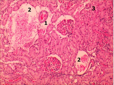
Results resource: Already after 1 day of the experiment there were changes related to the blood stream, particulary, in the arteries and veins there was an increased blood filling in comparison with the control rats.
Symbols: 1 – lobular artery, 2 – vein lumen with erythrocytes, 3 – arteriole.
Figure 1 – Histological section of a rat kidney after 1 day of being on an anhydrous diet. Staining with hematoxylin and eosin. ᵡ 140
More significant changes appeared already on the 3rd day of the anhydrous diet. The tone of the walls of partial and arcuate arteries was significantly increased and in the lumen of these vessels there was an accumulation of shaped elements. The changes in the lobular arteries were somewhat different: their lumen was expanded and the content of formed blood elements was significantly lower than in the previous generation. After the 6th day of dehydration the tone of partial arteries continued to increase and their lumen narrowed which was densely filled with formed elements. In the lobular arteries and arterioles the lumen was somewhat wider and the walls were thiner which indicated a decrease in the tone of the smooth muscle membrane. In the absence of water for 10 days the walls of partial arteries thickened and the lumen narrowed and was filled with shaped elements. The formation of thrombotic masses was noted in the veins that were nearby.
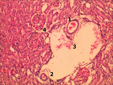
Symbols: 1 – lobular artery, 2 – arterioles, 3 – vein lumen, 4 – renal corpuscles and arteriole.
Figure 4.12 – Histological section of a rat kidney after 10 days of being on an anhydrous diet. Staining with hematoxylin and eosin. ᵡ 100
As for the small arteries they had thinned walls a widened lumen and only insignificant accumulations of formed elements that is dilatation reactions. Adjacent veins regardless of dilation were anemic.
With mild and moderate degrees respectively 3 rd and 6 th days of an anhydrous diet anincreased tone narrowing of the lumen of vessels and a decrease in the permeability of extra-organ and intra-organ arteries at the level of partial and arch arteries were noted. At the same time lobular arteries and arterioles acquired the opposite changes namely a tendency to vasodilation. With a severe degree of dehydration (10 days of total dehydration) the previous changes intensified except for the arcuate arteries where there was an inversion of the smooth muscle layer which led to a change in constriction reactions to dilation reactions.
Conclusions: Changes in the vascular bed of the kidneys depend on the degree of general dehydration. Сan be assumed the state of small vessels is necessary to maintain the blood supply of nephrons in the event of insufficient nutrition from the main vessels. The obtained data fully confirm the current ideas about dehydration as a cause of hypovolemia in which there are vascular reactions aimed at the centralization of blood circulation, which threatens hypoperfusion of internal organs, including the kidneys.
Reference:
1. Michael J Somers, MD Clinical assessment of hypovolemia (dehydration) in children https://www.uptodate.com/contents/clinical-assessment-of-hypovolemia-dehydration-in-children
2. Shanley L, Mittal V, Flores G. Preventing dehydration-related hospitalizations: a mixed-methods study of parents, inpatient attendings, and primary care physicians. Hosp Pediatr. 2013 Jul. 3(3):204-11.
|