Introduction. The discovery of the microorganism Aerococcus viridans is associated with the research of Williams, Hirch and Cowan. [1]. Deibel и Niven [2] noted their resemblance to Gaffkya homari, causative agents of infections in lobsters, and with pediococci contaminating canned meats and pickles. As a result of in-depth studies of the taxonomy of these microorganisms, it was proposed to preserve the genus Aerococcus [3]. The isolation of aerococci from the objects of the hospital environment is associated with their persistence in these conditions in view of their isolation from "liquid drugs" [4], from the lungs of a patient with suspected tuberculosis [5]. Aerococcus contamination of various items in the hospital environment has been shown. [6].
In 1965 Gorbunova M.L. [7] isolated cocci from breast milk that inhibit the growth of opportunistic cocci and rod-shaped bacteria. These microorganisms have been classified as Mycococcus hyperoxydans. As a result of the exclusion of the genus Mycococcus from the international classification [8], these microorganisms were reclassified to the genus "Aerococcus", species "viridans" [9]. It was found that aerococci produce hydrogen peroxide [10,11] as a result of the oxidation of lactic acid by NAD-independent lactate oxidase. [12].
Aerococci are widely distributed in the environment, where their reservoir is different types of living organisms, including them in the biotopes of their non-sterile shells. [13]. Based on their isolation from pathological foci, their role in aerococcal infections is discussed. Recently, the use of MALDI-TOF MS (Matrix Laser Desorption Ionization Time-of-Flight Mass Spectrometry) led to the identification of these isolates as aerococci. [14, 15].
The wide distribution of aerococci in the environment and in macroorganisms and their isolation from pathological material during infectious processes makes it possible to assume, on the one hand, the heterogeneity of their population [16], and on the other hand, a violation of the immune status of people with aerococcal infection. Risk factors for systemic A. viridans infections (bacteremia/endocarditis) are associated with: granulocytopenia, oral mucositis, prolonged hospitalization, prior antibiotic therapy, invasive procedures, foreign body implantation [17], which makes it possible to consider them as conditionally pathogenic bacteria.
The use of sophisticated instrumental methods for the identification of aerococci, such as (MALDI-TOF MS), is possible after the bacteriological method of isolating aerococci on standard nutrient media and their biochemical identification. We propose a preliminary stage that allows us to simplify and improve the accuracy in the isolation and identification of aerococci.
Materials and methods. For the isolation and preliminary identification of aerococci from the external environment and macroorganisms, the method was used [18]. For this, a selective indicator medium (IM) was used. [19]. The composition of the medium included (g per 1 liter of water): potassium iodide - 0.4, soluble starch - 10.0, nutrient agar - 30.0. Inoculation was performed and plates were incubated at 370C for 24-48 hours until colonies appeared. To account for the indicator reaction (IR), 5 ml of 10% H2SO4 was applied to the dish surface. After 3-5 minutes, the IR was recorded in the form of the appearance of dark purple zones around the growth of colonies. This method made it possible to quantitatively and qualitatively install the presence of colonies of microorganisms producing hydrogen peroxide.
To identify the indicator reaction of cultures without treatment of plates with H2SO4, the concentration of KJ was increased in the composition of IМ to 26 g per 1 liter. In the process of growth, colonies of aerococci on this medium turn dark purple without treatment with sulfuric acid, and it becomes possible to isolate pure cultures. This medium is effective in isolating aerococci from contaminated biological material. When using these two variants of media, the total oxidative activity (ox+) of aerococci was determined.
To identify the lactate oxidase activity of cells and cell-free components of aerococci, minimal indicator medium (MIМ) was used, in which distilled water after double purification was used instead of tap water, and highly purified agar-agar was used instead of standard nutrient agar.
Results. To isolate colonies of aerococci from contaminated biological material, an IМ with a high content of KJ was used, on which, without surface treatment of the medium with 10% H2SO4, dark purple colonies (ox+) appear among the colonies of associated microorganisms after 2-5 days, oxidizing KJ. (Fig. 1 ).
Fig. 1. Growth of ox+ colonies of A.viridans on IМ
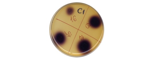
The oxidase activity of isolated pure cultures from these colonies was tested using the first version of the IS containing 0.4 g of potassium iodide per 1 liter of medium. Sowing of pure cultures was carried out, followed by treatment with sulfuric acid and taking into account the indicator reaction (Fig. 2).
Fig.2. IR on IМ around growth selected crops after treatment with H2SO4
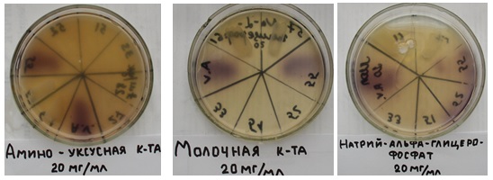
For preliminary selection of aerococci, putative oxidation substrates were added to the composition of the indicator medium: aminoacetic acid, sodium α-glycerophosphate, and lactic acid. For further work, cultures were selected that gave the most intense color (Fig. 3).
Fig.3. Change in the color of the indicator medium during the oxidation of the added
substrates.
Testing of lactate oxidase activity (LO) of selected pure cultures was carried out on minimal indicator medium (MIМ) with lactic acid. Isolated cultures were applied as plaques (Fig. 4)

Figure 4. IR of medium with lactic acid around plaques cultures with LO activity
The plaques of the cultures applied to the MIМ surface turned black as a result of the production of H2O2 during the oxidation of lactic acid. To identify the substrate for oxidation by isolated cultures, a suspension of washed cells was applied to the surface of the third medium variant (MIМ) in the form of a lawn, and the dishes were dried. Lactic acid at a concentration of 0.2 ml of 0.01% (LA) was added to the neutral cylinder in the center of the dish. For a certain time, a color reaction initiated by H2O2 developed, the diameter of which expanded as hydrogen peroxide diffused. Petri dishes show the reaction of the medium during the oxidation of lactic acid by the strain A.viridans 167 (Fig. 5,6,7).
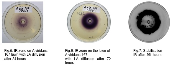
One can see the consistent development of the reaction to IS as LA diffuses and is oxidized by A.viridans cells.
There is a ring-shaped development of color with zones of its absence inside the diffusion of MK. This phenomenon can be explained by the change in oxidase activity to peroxidase activity when antioxidant mechanisms are switched on in certain ring zones. The formation of microcolonies was noted, due to the oxidation of lactic acid.
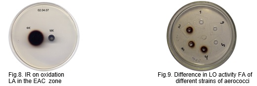
Lactate oxidase activity of enzymatic aerococcal complexes (EAC) was identified on a minimal indicator medium. EAC and lactic acid were introduced into the wells on the MIM surface. As a result of the diffusion of LA and its oxidation by EAC, an indicator reaction (IR) occurred. Fig/ (8) shows the staining zone around the cylinder with EAC (lactate oxidase), initiated by diffusing lactic acid (LA), and fig. (9) shows the indicator reaction around the wells with EAC from different strains of aerococci. During LA diffusion, an IR ring always forms around a cylinder containing EAC.
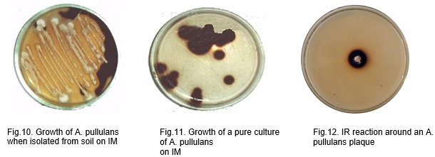
Nutrient indicator medium (IM) containing an increased concentration of KJ can be used to isolate and study the oxidizing ability of various microorganisms that produce hydrogen peroxide during the oxidation of biological substrates. Figures (10,11,12) show a photo of the reaction of the medium during the growth of Aureobasidium pullulans B5, isolated from the soil and having glucose oxidase activity [20]. When isolating the culture of A. pullulans B5, in order to detect the glucose oxidase activity of the colonies, glucose in an increased concentration (5–10%) was introduced into the composition of IM.
When the medium is contaminated from the air, cultures of microorganisms can grow on the IR using unidentifiable oxidation substrates contained in the nutrient medium.
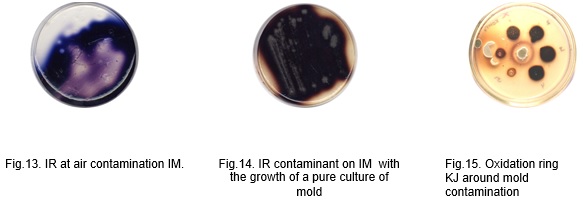
Figures (13,14,15) show dishes with the growth of mold microorganisms that initiate IR during their growth.
Conclusion. Thus, the proposed method for isolating microorganisms that oxidize certain substrates from the objects allows for preliminary screening, including the identification of specific activity. Having isolated strains with lactate oxidase activity and subsequently using biochemical, genotypic methods, including the method (MALDI-TOF MS), microorganisms of the A.viridans species are identified. The method makes it possible to drastically narrow the range of studied objects and improve the accuracy of identifying the oxidative activity of isolates.
Literature.
1. Williams, R.E.O., Hirch A., Cowan S.T. Aerococcus, a new bacterial genus. J. Gen. Microbiol. 1953. 8:475-480.
2. Deibel R.H., Niven C.F. Comparative study of Gaffkya homari, Aerococcus virias, tetrad-forming cocci from meat curing brines, and the genus Pediococcus. J. Bacteriol. 1960. 79:175-180.
3. Whittenbury R. A study of some pedio cocci and their relationship to Aerococcus viridans and the enterococci. J. Gen. Microbiol. 1965. 40:97-106.
4. Clausen 0.G. The discovery, isolation, and classification of various alpha-hemolytic micrococci which resemble aerococci. J. Gen. Microbiol. 1964.35:1-8.
5. Deibel R.H., Silliker J.H., Fagan P.T.. 522 Appl. Microbiol. A. viridans in the hospital environment. Some characteristics of an oleate-requiring hemolytic Pediococcus. J. Bacteriol. 1964.88: 1078-1083.
6. Mildred A., Kerbau G.H., Evans J B. Aerococcus viridans in the Hospital Environment. Appl.microbiology, Mar. 1968, Vol. 16, No. 3. 519-523.
7. Горбунова М.Л. О группе бактерий, продуцентов перекисей, обитающих в организме человека // Антибиотики. - К., 1965. - №1. - С.191-194.
8. Evans, J. B. 1986. Genus Aerococcus Williams, Hirch and Cowan 1953, 475AL, p. 1080. In P. H. A. Sneath, N. S. Mair, M. E. Sharpe, and J. G. Holt (ed.), Bergey's Manual of Systematic Bacteriology, vol. 2. The Williams & Wilkins Co.Baltimore.
9. Горбунова М.Л., Кременчуцкий Г.Н. Физиологические и биохимические механизмы активности микроорганизмов рода Mycococcus (Aerococcus) /В кн.: Матер. УII съезда Всесоюз. о-ва микробиологов, 1985. - Алма-Ата, 1985. - Т.1. - С.14.
10. Кременчуцкий Г.Н., Самойленко И.И. Действие перекиси водорода, продуцируемой Aerococcus viridans, на E.coli, B.subtilis // Микробиол. журн. - 1987. - Т.49, № 2. - С.91-93.
11. Кременчуцкий Г.Н. Сравнительная оценка антагонистически активных форм кислорода физического и биологического происхождения[Текст]/ Г.Н.Кременчуцкий, Л.Г.Юргель, А.В.Шарун// ХII Международная конференция и дискуссионный научный клуб "Новые информационные технологии в медицине и экологии": Крым, Ялта-Гурзуф.-2004.- С. 180.
12. Кременчуцкий Г.Н., Аренков П.Я. Лактатоксидазная активность культур Aerococcus viridans[Текст]/ Г.Н.Кременчуцкий, П.Я. Аренков //Микробиол.ж.-1989.-Т.5;№5.-С.17-20.
13. Микроэкология аэрококков и проблемы их использования в качестве пробиотических препаратов / Д.О. Степанський, Г.Н. Кременчуцкий, Л.Г. Юргель, О.В. Шарун [и др.]/ Труді ХХ междунар. конф. и дискуссионного науч. клуба «Новые информационные технологии в медицине, биологии, фармакологии и экологии». – Ялта, Гурзуф, 2012. – С. 72- 74.
14. Rasmussen M. Aerococcus: An increasingly acknowledged human pathogen. Clin Microbiol Infect. 2016;22:22–27. [PubMed] [Google Scholar] ; Senneby E, Nilson B, Petersson AC, Rasmussen M. Matrix-assisted laser desorption ionization-time of flight mass spectrometry is a sensitive and specific method for identification of aerococci. J Clin Microbiol. 2013;51(4):1303–04. [PMC free article] [PubMed] [Google Scholar].
15. Senneby E, Göransson L, Weiber S, Rasmussen M.A. Population-based study of aerococcal bacteraemia in the MALDI-TOF MS-era. Eur J Clin Microbiol Infect Dis. 2016;35(5):755–62. [PubMed] [Google Scholar]
16. Гетерогенність бактерій роду Aerococcus та її роль в розробці нових пробіотиків і контролю їх автентичності . 2002 г. Автореф. дис... канд. мед. наук: 03.00.07 / С.А. Черняєв; АМН України. Ін-т мікробіології та імунології ім. І.І.Мечникова. — Х., 2002. — 22 с. — укp.
17. Uh Y, Son JS, Jang IH, Yoon KJ, Hong SK. Penicillin-resistant Aerococcus viridans bacteremia associated with granulocytopenia. J Korean Med Sci 2002;17:113-5.
18. Кременчуцкий Г.Н., Горбунова М.Л., Маганчук В.П. Методика выделения и идентифика-ции гетеротрофных бактерий, продуцирующих перекись водорода // Информационное письмо. Киев: МЗ УССР, 1987.- 2с.)
19. Kremenchutsky G.N. AS №1494516 USSR. Nutrition medium for determinatiom of lactate-oxidative activity of Aerococcus viridans / Publ. in BJ 1989, № 2610.
20. Смотрова Н.Г. Выделение штаммов микроорганизмов, обладающих глюкозооксидазной активностью, из окружающей среды [Текст] / Н. Г. Смотрова, Г. Н. Кременчуцкий //Мікробіол. журн. - 2002. - T64; № 6. - С. 28-34.
|