Abstract
The article is devoted to the urgent problem of objectification of small vessel vasculitis at the level of the microcirculatory bed, visual verification in vivo of the structural transformation of capillary blood filling and blood flow in the superficial and deep layers of the microcirculatory bed. The term vasculitis has long considered vascular pathology as an inflammatory process.
Vasculitis is a heterogeneous group of diseases characterized by inflammation of the vacular wall of various calibers. The clinical signs of vasculitis depend on the type of affected vessels, involved organs and systems, and the activity of the inflammatory process, and can vary from local skin symptoms to life-threatening systemic lesions. The article considers the main clinical signs of vasculitis depending on the classification and caliber of the affected vessels. The authors have analyzed possibilities of assessing the state of capillaries and differentiating different forms of vasculitis thanks to vascular screening technology. They’ve considered some aspects of indirect signs of damage to the vascular wall of arterioles, venules and capillaries with disorders of blood filling and blood flow in various segments of the microcirculatory bed and established specific patterns of changes in microangioarchitectonics in the rheumatological spectrum of damage to the microcirculatory bed.
Keywords: vasculitis, small vessel vasculitis, clinical manifestations, ANCA vasculitis, cutaneous purpura, glomerulonephritis, systemic vascular disease, vascular screening technology, rheumatic capillary disease, Raynaud's syndrome, lupus erythematosus, systemic scleroderma, Covid, blood supply, capillaries, blood supply deficiency, microcirculation, rheumatic vasculitis, capillaries, venular stasis.
Introduction
Vasculitis is a disease in which there is an immunoinflammatory lesion of the walls of blood vessels. Depending on the caliber of the affected vessels, vasculitis is divided into large-caliber, medium-caliber and small-caliber. This division determines the clinical picture of the disease, because the lesion of large vessels leads to ischemia of large organs, while small-caliber vasculitis more often has predominantly cutaneous or nephrological manifestations. Vasculitis can be primary (idiopathic) or secondary, caused by infection, neoplasms or other autoimmune processes.
At the same time, intravital visualization of capillaries thanks to the AngioVeritasR vascular screening technology applying optical smart wide-format capillaroscopy has enables us to take a new look at capillary lesions in real life and partially change our views on the specific pathohemodynamic patterns of microcirculation disorders in vasculitis.
The relevance of vasculitis of various etiologies, including rheumatoid vasculitis
Rheumatic diseases is a group of diseases of the connective tissue (dermis, tendon-ligamentous apparatus, cartilage, bone tissue, synovial and serous membranes, basement membranes, vessels, etc.) in the pathogenesis of which the main role is played by multisystem autoimmunity in response and the inflammatory process associated with symptoms from many organs and systems.
One of the key links in this process is the vascular system, which sooner or later is involved in the process, and perhaps plays a major role in the development and progression of rheumatic diseases. From the position of the basics of hemodynamics, the blood supply to the extremities occurs according to the residual principle, with a background diastolic deficiency of blood supply in comparison with full hemodynamics in the main arteries of vital organs and systems. At the same time, the blood supply to muscles, bones, connective tissue formations, joints and bones occurs practically according to the peripheral type with a minimum of background blood filling of capillaries. Therefore, the study of the microcirculatory bed in the nail bed of the fingers of the upper and lower extremities plays a decisive role in the visualization of microcirculatory disorders and in the diagnosis of vasculitis [1].
The term rheumatic vasculitis has long existed as a sign of rheumatic vascular damage, but for the most part it has a general nature and is often used as a concomitant addition to the diagnosis.
In fact, there are many scientific works related to the study of vessels in rheumatic diseases and the search for specific patterns - thickening of the intima-media complex and vascular wall in collagenoses, description of capillaroscopic patterns in rheumatology, etc.
However, there is no systematic approach to pathological restructuring of the cardiovascular system in rheumatological diseases, explanation of the influence of pathological transformations of vessels on the general state of perfusion in the body and search for ways to overcome local dyshemias with the aim of sanogenic restoration of the body.
With the advent of modern methods of vascular visualization, it became possible to take a new look at changes in the microvascular bed that are specific to Raynaud's syndrome, systemic lupus erythematosus, systemic scleroderma, uncontrolled neoangioncogenesis.
A term rheumatic vasculitis has been known for a long time and often accompanies the description of the clinic of rheumatism as a collective term without clear criteria for its identification and etiopathogenesis. Historically, vasculitis was theoretically justified by the process of inflammation of the vascular wall (infectious-allergic and autoimmune genesis), which accompanies rheumatological diseases.
Angiotrophoneurosis, hemorrhagic vasculitis, capillary toxicosis, Raynaud's syndrome etc.are used to characterize rheumatic vasculitis [2,3].
At the same time, there are no clear criteria for certain types of rheumatic vasculitis that would have an evidential applied nature and clear patterns of their definition.
The axiom of inflammatory vascular wall damage under the term vasculitis still prevails, which requires confirmation or refutation by native visualization of processes occurring at the microcirculatory level.
Let's briefly classify vasculitis:
1. Common symptoms of vasculitis
Many patients with vasculitis have nonspecific first signs, similar to a systemic inflammatory syndrome:
• fever;
• weakness, general asthenia;
• weight loss;
• arthralgias or arthritis;
• anemia and increased ESR/CRP;
• skin rashes.
Such symptoms often precede organ damage and can be misleading, which complicates early diagnosis.
2. Clinical signs depending on the caliber of vessels
2.1. Large-caliber vasculitis
They include giant cell (temporal) arteritis and Takayasu arteritis.
Giant cell arteritis:
• headache, mainly in the temporal region;
• pain when chewing (claudication of the jaws);
• decreased vision, up to blindness;
• tenderness of the temporal artery on palpation.
Takayasu's arteritis:
• lack of pulses in the extremities;
• difference in blood pressure in the arms;
• murmur over the large arteries;
• symptoms of ischemia of the extremities and internal organs.
2.2. Medium-sized vasculitis
Polyarteritis nodosa (PNA) is a classic example
Clinical signs of PNA:
• fever, weight loss, general weakness;
• myalgias and arthralgias;
• kidney damage (arterial hypertension, proteinuria, hematuria);
• abdominal pain, gastrointestinal bleeding;
• peripheral nerve damage (mononeuropathy);
• skin nodules, livedo reticularis.
2.3. Small-caliber vasculitis
This group includes ANCA-associated vasculitides and immune complex vasculitides.
Anka-associated vasculitis:
• Granulomatosis with polyangiitis (formerly Wegener's disease):
o chronic rhinitis, nasal obstruction;
o mucosal ulcers;
o glomerulonephritis;
o pulmonary infiltrates, hemoptysis;
o middle ear lesions.
• Microscopic polyangiitis:
o renopulmonary syndrome;
o rapidly progressive glomerulonephritis;
o shortness of breath, cough, alveolar hemorrhage.
• Eosinophilic granulomatosis with polyangiitis (Churg-Strauss syndrome):
o asthma;
o eosinophilia;
o polyneuropathies;
o skin rashes;
o heart disease (pericarditis, myocarditis).
Immune complex vasculitis:
• Henoch-Schönlein purpura (IgA-vasculitis):
o palpable purpura on the lower extremities;
o arthralgias;
o abdominal pain;
o nephritis (proteinuria, hematuria).
• Cryoglobulinemic vasculitis:
o purpura, edema;
o peripheral neuropathy;
o hepatitis C in anamnesis;
o renal syndrome.
3. Cutaneous manifestations of vasculitis
The skin is one of the most common target organs in vasculitis. Typical clinical features:
• palpable purpura (especially in small-caliber vasculitis);
• necrotic lesions;
• nodules;
• livedo reticularis;
• urticarial vasculitis;
• gangrene of fingers.
It should be emphasized that changes in the blood supply to the capillary bed in the form of venular stasis and thrombosis in the superficial layers of the dermis are accompanied by visualization of the vascular pattern of superficially located small vessels and capillaries, which previously allowed one to suspect a particular nosology based on the vascular pattern on the skin surface.
4. Neurological complications
Peripheral nervous system involvement most commonly occurs in peripheral arterial vasculitis (PAV), Churg-Strauss vasculitis, and cryoglobulinemic vasculitis.
Sign:
• mononeuritis;
• sensory-motor polyneuropathy;
• less often – strokes in case of damage to cerebral vessels.
In this situation, the main role in the damage to peripheral nerves is played by the vasa vasorum around the nerve trunks.
Regarding the cerebral arteries, the diagnosis of rheumatic vasculitis of the cerebral arteries remains relevant.
5. Cardiovascular lesions
May be shown by:
• arterial hypertension (renovascular origin) and venous hypertension;
• myocardial ischemia;
• pericarditis, myocarditis;
• aneurysms (especially in Kawasaki disease).
6. Pulmonary signs of vasculitis
More common in ANCA-associated vasculitis (associated with the presence of antibodies to neutrophil cytoplasmic antigens (ANCA):
• cough;
• shortness of breath;
• hemoptysis;
• interstitial changes on CT;
• alveolar hemorrhage.
Thus, vasculitis is a complex clinical nosology with diverse signs that cover almost all organs and systems. Important for diagnosis is the assessment of the nature of skin changes, lesions of the respiratory and urinary systems, the presence of systemic symptoms and typical laboratory changes (ANCA, IgA, cryoglobulins, etc.).
Early recognition of clinical manifestations reduces the risk of disability and improves the prognosis of patients [4,5].
Modern diagnostic equipment enables for live visualization of the vascular bed both at the level of main arteries and veins, and at the level of peripheral and terminal segments of arteries and veins - arterioles and venules. Live visualization of the microcirculatory bed enables for verification of vascular disorders and tangential assessment of structural changes in the capillary profile, clearly identifying disorders of blood supply in the arteriolar and/or venular beds (AngioVeritasR vascular screening technology).
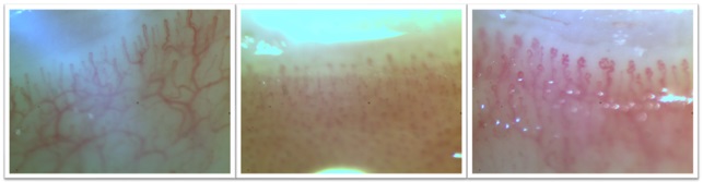
Figure. 1 Image of the capillary bed in vasculitis
The presented images show different degrees of blood filling and blood flow disorders in classical capillaries and in structurally altered capillaries of the superficial and deep layers of the microcirculatory bed. It should be emphasized that the first signs of microcirculatory changes can precede laboratory verification by six months to a year.
On the other hand, visualization of the state of changes in the microcirculatory bed thanks to wide-format optical digital vascular screening technology allows pathophysiological detection, modeling and correction of vascular bed disorders according to the lesion link - capillaries, arterioles, venules and perivascular edema, leading to a visible deficiency of blood filling and blood supply.
The capillary visualization method is highly informative, highly sensitive to structural and functional rearrangements of the microcirculatory bed - sensitivity 98% for various microcirculatory disorders, specificity for rheumatic vasculitis - 95%.
The question of hemodynamic models in vasculitis disorders naturally arises, since the optical imaging method is based on the principle of visualizing blood filling and blood flow, and the walls of capillaries and small vessels themselves are not visualized.
From the standpoint of the laws of hemodynamics in general and in the microcirculatory bed, the proportion between the calibers of the arteriolar and venular segments of capillaries and the transitional elbow should be preserved.
The situation when the arteriolar segment in the proximal segment is not filled with blood and was previously considered as arteriolar spasm, today does not correspond to the hemodynamic parameters of arteriolar spasm, and most likely is a sing of edema of the capillary wall with a sharp thinning of the proximal arteriolar segment of capillaries, which causes pallor of the skin of the fingers of the extremities. At the same time, the expansion of the venular segment and venular stasis demonstrate a cyanotic shade of the skin color of the fingers of the extremities.
According ot mathematical modelling of hemodynamic disorders, one should take into account the preservation of the classical shape of the capillary or its structural changes, the presence of post-COVID microthromboangiopathy of varying severity, the magnitude and nature of perivascular edema, the patterns of pathological structural transformations of the surface layer of capillaries in rheumatic vasculitis - Raynaud's syndrome, systemic scleroderma, lupus erythematosus, rheumatoid arthritis, etc. All these nosologies have specific patterns of microcirculation disorders, which require an individual approach to vascular therapy (Angiotherapy and Angiocorrection of arteriovenular balance at the microcirculatory level) along with specific treatment of the underlying disease of the rheumatological spectrum.
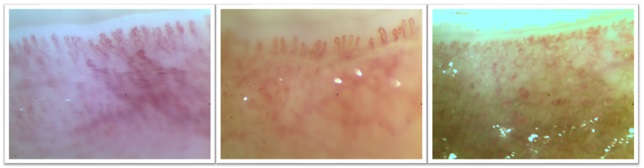
Figure 2. Specific patterns of rheumatic vasculitis in different nosologies.
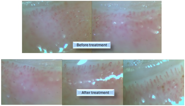
Figure 3. Hemodynamic correction of rheumatic vasculitis aimed at restoring the physiological characteristics of microangioarchitectonics and blood flow.
Historically, rheumatic diseases have been considered as infectious, autoimmune diseases with a corresponding direction of treatment of the etiopathogenetic factor.
Anti-inflammatory treatment is complex and practically does not reach the stage of cure and recovery. Therefore, diseases of the rheumatic spectrum remain not fully understood and require further research to establish all pathogenetic factors.
On the other hand, rheumatic vasculitis are increasingly common in medical practice. Therefore, studies of vascular changes and disorders in vivo require in-depth research and the search for effective ways to overcome pathological hemodynamic patterns, expansion and detailing of hemodynamic pathological patterns in the diagnostic protocol of the vascular arteriovenous status of the regional limbic reservoir of the upper and lower extremities in vivo, both by ultrasound at the macroangiological level and by optical capillaroscopy at the microangiological level using digital wide-format optical vascular screening technology.
Over the past decade, specific patterns of changes in the shape of nail bed capillaries have been identified in patients with the rheumatological spectrum, which are highly informative, highly differentiated in systemic scleroderma and lupus erythematosus, and today in fact belong to nosologically specific patterns.
However, these patterns of giant capillaries, capillary hematomas, avascular zones, and pathological neoangiogenesis in the transitional knee have not been scientifically substantiated with regard to the etiopathogenetic and hemodynamic prerequisites for changing the shape of capillaries and pathological hemodynamic rearrangements in affected capillaries up to complete cessation of blood flow, differentiation of the phenomenon of shadow capillary desolation, and the rheumatological mechanism of the formation of post-hematoma avascular zones.
Angiocorrection and angiotherapy of blood filling and blood flow disorders in patients of the rheumatological spectrum based on evidence-based medicine involves quantitative and qualitative analysis of existing microcirculation disorders, modelling of various micro- and macrohemodynamic situations with the applied application of the laws of hemodynamics in order to restore the structure and function of the vascular wall of capillaries for blood mass transfer, sanogenic transformations in structurally provoked capillaries, reduction of perivascular edema, reduction and leveling of blood filling deficiency of the capillary bed, reduction of venular stasis, restoration of pulse wave and blood flow in capillaries.
Client-oriented Angiocorrection of microthromboangiopathy, capillary hematomas, giant capillaries and pathological neoangiogenesis (neoangionecogenesis) involves the detection of single pathologically altered capillaries (this requires a large-format image with visualization of about 100 capillaries). Even the presence of 1 pathological capillary in the field of view of 100 capillaries indicates early diagnosis of the process and the possibility of its hemodynamic correction. When visualizing 3-5 capillaries with a training finger microscope and not detecting single pathological capillaries is regarded as a technical limitation of the device - a finger microscope and a technically determined medical error.
The peculiarity of pathological forms of capillaries of the rheumatological spectrum lies in the critical state of blood flow and the urgent need for client-oriented Angiocorrection, since pathological changes in blood flow occur: both functional hemodynamic and structural pathological rearrangements of the microcirculatory bed, which are different in nature individually for each patient.
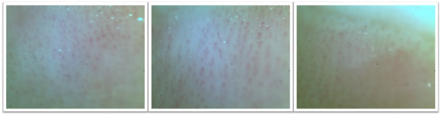
The state of microcirculation has significantly worsened during the Covid-19 pandemic and has been complicated by expressed signs of microthromboangiopathy, often of an occlusive nature with a perfusion block in the entire field of capillaries of the microcirculatory bed.
Figure 4. Post-COVID microthromboangiopathy in the deep layers of the microcirculatory bed (AngioVeritasR vascular screening technology)
These pathological changes that became the object of our study in order to find hemodynamically justified angiocorrective algorithms for restoring an adequate level of blood supply to the hands and feet of patients, vascularization of the skin, which significantly deteriorated after suffering from Covid or vaccination against Covid, which led to the search for adequate angiocorrective effects on the sanogenic restoration of blood filling and blood flow, vascular tube patency, the existing critical picture of microcirculation and the absence of perfusion in Long Covid. Occlusive microthromboangiopathy has become a new challenge for the medical scientific community in the search for new hemodynamic algorithms for restoring blood supply in the deep layers of the microcirculatory bed.
Conclusions
Vasculitis is a complex clinical nosology with a variety of signs that cover almost all organs and systems.
Early recognition of clinical sign enables to reduce the risk of vascular blood flow disorders, mortality and disability, improving treatment and improving the prognosis of patients.
Thus, today vasculitis can be objectified from the standpoint of hemodynamic blood supply disorders, visualized live and conduct complex infection-specific therapy along with vascular therapy with monitoring of sanogenic changes in the blood microcirculation.
In the 5 years since the beginning of the Covid-19 pandemic, world civilization has faced a new challenge - post-Covid blood supply disorders, when vascular disorders have moved from the category of non-infectious diseases to complex chronic post-infectious microthromboangiopathy in the deep layers of the microcirculatory bed and high risks of pathological vascular transformations of the rheumatological and/or oncological spectrums.
References
1. Lushchyk UB, Novytskyy VV et al. (2021). Vascular Screening of PathoNeoAngioOncogenesis. (Analytical Approach to an Early Diagnosis of Pathological ArterioVenous Angiotransformations at the Micro- and Macrovascular Levels). Journal of Blood Disorders, Symptoms & Treatments- 2021- Vol. 3
2. Abul-Fadl, A.M.A.M.; Mourad, M.M.; Ghamrawy, A.; Sarhan, A.E. Trends in Deaths from Rheumatic Heart Disease in the Eastern Mediterranean Region: Burden and Challenges. J. Cardiovasc. Dev. Dis. 2018, 5, 32. https://doi.org/10.3390/jcdd5020032
3. Bekaryssova, D., Yessirkepov, M. & Mahmudov, K. Structure, demography, and medico-social characteristics of articular syndrome in rheumatic diseases: a retrospective monocentric analysis of 2019–2021 data. Rheumatol Int 43, 2057–2064 (2023). https://doi.org/10.1007/s00296-023-05435-x
4. Doornum, Sharon & Bohensky, Megan & Tacey, Mark & Brand, Caroline & Sundararajan, Vijaya & Wicks, Ian. (2015). Increased 30-day and 1-year mortality rates and lower coronary revascularisation rates following acute myocardial infarction in patients with autoimmune rheumatic disease. Arthritis research & therapy. 17. 552. 10.1186/s13075-015-0552-2.
5. P.-C. Wu, M.-N. Huang, Y.-M. Kuo, S.-C. Hsieh, C.-L. Yu. Clinical applicability of quantitative nailfold capillaroscopy in differential diagnosis of connective tissue diseases with Raynaud's phenomenon. J Formos Med Assoc, 112 (2013), pp. 482-488 http://dx.doi.org/10.1016/j.jfma.2012.02.029
|